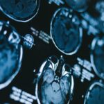
Digital x-ray
Traditional x-rays capture images from the body onto films. This helps doctors find abnormal structures that they may not otherwise be able to feel when they examine the patient. Digital x-rays offer two distinct advantages. Firstly, as digitally-captured images, they are available for immediate viewing without the lengthy and complex steps of chemical processing of films. Secondly, they use and therefore expose you to much less radiation.
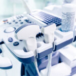
3D ultrasound scan
Ultrasound uses sound waves to create pictures of internal organs and tissue and detect any changes. In the diagnosis of some cancers, like breast cancer, it can help distinguish possible tumours from harmless cysts. While 2D ultrasound only gives outlines and flat-looking images, the more modern technology of 3D ultrasound provides a more realistic view of the body, particularly of the internal organs.
3D mammography
A 3D mammogram uses low-dose X-rays to create a three-dimensional image of the breast. It is useful for breast cancer screening tests and detecting abnormalities in patients with no symptoms. It can also be used to investigate problems like lumps, nipple discharge or changes in breast shape.
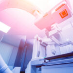
CT scan
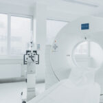
To analyse changes in any part of the body, CT scans generate a combination of three-dimensional images and powerful, digital X-rays. Together, these two imaging techniques will provide a detailed picture of a tumour’s location, shape and size, and any changes over time.
CT-simulation machines are used to accurately plan the delivery of radiation treatment to treat cancer cells in the safest way.
Biopsies
When diagnostic imaging reveals an abnormality such as a lump, further investigation is usually required to determine whether the lump contains cancer cells – and if so, to give further information on the type and extent of the cancer.
In a biopsy, a small sample of the tissue is removed and examined under a microscope. The test will show whether the tissue is benign and harmless, or malignant (cancerous) and likely to spread.
Blood tests
Marcelle Ruth has automated laboratory facilities which provide a wide-range of blood tests including monitoring of tumour markers: substances made by either cancerous cells or healthy cells that are fighting cancer. As tumour markers can show up in benign conditions, they are not used to diagnose cancer. But after a positive diagnosis, monitoring marker levels can show whether a treatment is working or a cancer is spreading.